Speaking Valve Use During Mechanical Ventilation: More than Just for Communication and Swallowing
Gail M. Sudderth, RRT

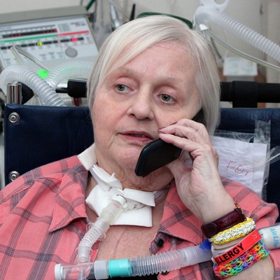
The inability to communicate during periods of mechanical ventilation (MV) can increase psychoemotional distress (Egbers, Bultsma, Middlekamp, & Beoerma, 2014) and has been associated with depression and post-traumatic stress disorder (Freeman -Sanderson, Togher, Elkins & Phipps, 2016). One way speaking valves can be used to restore verbal communication for patients who require MV. The Passy Muir® Valve is the only bias-closed position valve that can be used during MV. The Passy Muir Valve opens during inspiration and closes at the end of inspiration, re-directing exhalation through the vocal cords and out through the mouth and nose, which allows for verbal communication. The restoration of airflow, sensation, and positive airway pressure to the aerodigestive tract returns the upper airway to a more normal physiologic condition and may also have other clinical benefits for the patient who requires tracheostomy and MV.
It is common to delay intervention by speech-language pathologists and the use of speaking valves in the ICU on patients who require mechanical ventilation, based on the rationale that patients are “too sick.” The literature suggests that this hands-off approach may cause more harm than good and early intervention can minimize or potentially reverse the impact (Burkhead, 2011). The Speech-Language Pathologist (SLP)-Respiratory Care Practitioner (RCP) team is presented with a unique opportunity to co-treat patients who require tracheostomy and MV to provide not only a way to communicate, but a way to restore airflow and engage the glottis and restore positive pressure to the aero digestive tract. This therapy may enhance weaning and rehabilitation including safer swallowing to reduce aspiration (Amathieu et al., 2012; Rodrigues et al., 2015), improve swallow and cough (Pitts, et al., 2009), reduce respiratory infections (Carmona, Díaz, Alonso, Guarasa, Martínez, & López, 2015), promote alveolar recruitment (Sutt, Cornwell, Mullany, Kinneally & Fraser, 2015) and enhance early mobilization efforts (Massery, 2014).
With few exceptions, patients who require tracheotomy were previously intubated. The presence of an endotracheal tube, along with infection, medications, immobility, disuse atrophy, and co-morbidities often lead to ICU acquired muscle weakness. Muscle weakness is a factor in dysphagia and is associated with increased symptomatic aspiration risk leading to significant morbidity and mortality in ICU patients (Mirzakhani et al., 2013). Lack of airflow to the upper airway during endotracheal intubation continues after a tracheostomy tube is placed with inflated cuff and can lead to sensory changes in the mucosa of the oropharynx and larynx contributing to dysphagia (Burkhead, 2011). A significant number of these patients also develop diaphragmatic weakness as well, resulting in significantly longer duration of MV (Supinski & Callahan, 2012). Diaphragmatic force generating capacity may be reduced as much as 32% after just 5 or 6 days of MV (Schellekens et al., 2016). Respiratory weakness is often associated with difficult weaning and increased mortality. Therefore, it would be reasonable to consider Respiratory Muscle Training (RMT) as part of the weaning and rehab process, along with consideration for preventative strategies to reduce or slow disuse atrophy of the respiratory muscles (Schellekens et al., 2016). RMT has also been linked to improved swallow and cough (Pitts et al., 2009). RMT is generally defined as a technique for improving respiratory muscle function and includes performing inspiratory and/or expiratory maneuvers against a resistance. However, further research is needed to establish efficacy in certain patient groups and specific training protocols should be implemented for Respiratory Muscle Training specific to various patient populations. Inspiratory Muscle Training (IMT) has been reported to increase exercise endurance, muscle strength, and perceived dyspnea in patients with COPD (Geddes, O’Brien, Reid , Brooks & Crowe, 2008), and Expiratory Muscle Training (EMT) has been linked to reduced perception of dyspnea in COPD patients during exercise and improved cough and swallow safety (Laciuga, Rosenbek, Davenport, & Sapienza, 2014).
While patients with tracheostomy and MV participate in a weaning and rehabilitation process, they also must have their communication needs met. Clinicians would agree that patients may be able to employ different methods of verbal communication at varying times during their illness, and any and all methods of providing voicing should be explored. However, while some methods of ventilator assisted speech do assist the patient with voicing and the ability to communicate (McGrath, Lynch, Wilson, Nicholson & Wallace, 2016; Hoit, Banzett, Lohmeier, Hixon, & Brown, 2003), they do little to restore upper airway physiology to a more normal condition.
Some have suggested that partial cuff deflation during MV is a preferred means to accomplish speech; however, while this may be useful in a select group of patients who are unable to manage full cuff deflation, it may not be the best way to restore upper airway physiology. It was suggested by Hoit et al., (2003) that the combination of increasing inspiratory time and increasing PEEP, as high as 15 cmH2O in some subjects, produced a quality of voicing identical to using a speaking valve. The author also stated that “high PEEP is a safer alternative than a one-way speaking valve” (Hoit et al., 2003). However, it may be more likely that the subjects were performing high flow leak speech. High inspiratory flows, along with increased PEEP, may be difficult for weak patients to manage, leading to increased work of breathing, and/or breath stacking. It should be noted the authors did not have findings within the study to support the claim of improved safety with this method. In addition, encouraging speech while the ventilator is delivering an inspiratory breath is not natural speech, as natural voicing occurs during the expiratory cycle.
McGrath, Lynch, Wilson, Nicholson, and Wallace (2016) proposed an alternative method of ventilator assisted communication by using a tracheostomy tube with a subglottic suction port. The port is used to deliver a low flow of gas above the cuff, which may be inflated or partially deflated. The reported limitations to this method include limited voice quality, possible laryngeal injury with higher flows, stoma leakage of gas, and the dry gas delivery causing drying of the mucosa and hyper-adduction of the vocal folds (McGrath et al., 2016). While this method has its drawbacks, it may be a good alternative for the ICU patient who is too sick or unable to manage cuff deflation even for short periods of time. However, another consideration is that this is a specialized trach and may require a trach change for the patient.
While therapies like RMT assist with improving coughing, swallowing, and trunk strength, tasks such as walking, balance and exercise require engaging the glottis and airflow to the upper airway (Massery, 2014). Normalized voicing also requires engagement of the glottis and airflow through the upper airway. To achieve this engagement, the cuff must be completely deflated and a no-leak speaking valve placed on the tracheostomy tube to allow for 100% of exhalation to flow through the glottis, upper airway, mouth and nose. It is also important to understand how to maintain adequate ventilation with the cuff of the tracheostomy tube deflated. A thorough upper airway assessment to assure upper airway patency must be performed prior to use of a no-leak speaking valve. Some practitioners may be hesitant to try managing MV in the cuff deflation condition, concerned that adequate ventilation cannot be maintained. In a study done on “unweanable” ventilator dependent patients with neuromuscular disease, Bach reported that 91 out of 104 patients were adequately ventilated with either the cuff deflated or with cuffless tracheostomy tubes (Bach & Alba, 1990).
The most likely patient to manage cuff deflation is one who is medically stable, awake, and engages the voice. It might be appropriate to begin cuff deflation sessions in conjunction with sedation vacations (when sedating medications are not being used). The clinician should understand that a patient who has not felt airflow through the upper airway for several weeks, or even longer, may not achieve full cuff deflation in one session. Some ventilator adjustments that may make cuff deflation more successful include reducing or eliminating PEEP and/or changing sensitivity settings, so that the ventilator does not auto cycle. Sutt, Cornwell, Mullany, Kinneally, and Fraser (2016) reported improved lung recruitment when using the Passy Muir® speaking valve in conjunction with MV with the PEEP reduced or turned to zero. This improvement was maintained for a period of time, even after the one-way valve was removed. The authors attribute this maintenance to the return of a more normal upper airway resistance since exhalation occurred through the larynx and upper airway. At this stage of assessment, it is very important for the SLP and RCP to work closely together and employ strategies to assist the patient in maintaining adequate ventilation. The RCP will manage the ventilator alarms and monitor ventilation, while the SLP can cue the patient to breathe in during the inspiratory cycle of the ventilator and perform an expiratory maneuver to trigger the ventilator into exhalation in the presence of the leak during cuff deflation. This coordination with the ventilator is then transitioned to coordinating respirations with voicing on exhalation and may lead to coordinating respirations and swallowing. In addition to ventilator adjustments, the process of cuff deflation should not be rushed. Some patients will take longer to manage this step due to weakness of the laryngeal and pharyngeal muscles/structures and reduced sensation. A patient may exhibit coughing, throat clearing, shortness of breath, and other signs of adjustment – all of which are a part of the process in learning to coordinate breathing with the ventilator and developing a sense of normalcy with a return of airflow through the upper airway. Additionally, good oral care and suctioning as needed are important before and during this step of the airway assessment.
Once the cuff is completely deflated, airway patency can be determined by assessing voicing on exhalation, listening for exhalation though the upper airway using a stethoscope, or by reading the peak inspiratory pressure (PIP) and/or exhaled volumes via the ventilator. The clinician can objectively document an adequate leak and upper airway patency when reading a 40-50 percent drop in PIP and/or decrease in exhaled tidal volume measured by the ventilator. These measurements would suggest that the tracheostomy tube is properly sized to allow for sufficient airflow around the tracheostomy and upwards to the upper airway. It also suggests that there is no significant obstruction above the tracheostomy tube. A no-leak speaking valve then can be placed into the ventilator circuit while mechanical ventilation continues.
Once the no-leak valve has been placed in the ventilator circuit, the RCP and SLP continue to work together to assure patient-ventilator synchrony and adequate ventilation. The SLP may provide inspiratory and expiratory cues to the patient while the RCP monitors ventilation by monitoring PIP. PIP should be closely monitored since it is the measure of adequate ventilation comparable to pre-cuff deflation and no exhaled air will return or be measured by the ventilator. It may be necessary to increase delivered volume to achieve pre-cuff deflation PIP and assure adequate alveolar ventilation; however, this step may not be needed once the patient gets stronger. At this stage, the RCP should manage the ventilator alarm settings following safe practice. Other vent specific strategies may also be utilized depending on the mode of ventilation or brand, including flow or time limiting pressure delivered breaths and consider whether it is appropriate to use leak compensation as provided by the specific ventilator.
As the aerodigestive system is returned to the more normal condition with the use of a Passy Muir Valve in-line with MV, therapies that require glottis engagement, positive sub-glottic pressure, and airflow can begin. As previously mentioned, oral intubation and reduced airflow to the airway may result in decreased sensation, in addition to disuse atrophy and muscle weakness (Mirzakhani et al., 2013). Individualized therapeutic programs may be developed, requiring that therapies be modified for each patient dependent on the level of function. Progress may be slow in some patients with multiple co-morbidities but should be pursued when medically appropriate to ameliorate deterioration as much as possible.
One way speaking valves have long been used to allow for airflow through the upper airway for speech. Clinicians should consider the other possible benefits to the patient when airflow, sensation, and positive pressure is restored to the upper airway as part of the weaning strategy for patients who require MV. Use of a Passy Muir Valve during MV also provides improved access for treatment of dysphagia and increases participation in physical therapy through improved trunk stability and postural control, which may lead to improved weaning rates, reduced time of MV, and fshortened ICU length of stays. According to Grosu et al. (2012), (“Difficulties in discontinuing MV are encountered in 20% to 25% of patients who receive MV, with a staggering 40% of the time spent in the ICU devoted to weaning from MV. Hence, techniques that expedite the weaning process should have a profound effect on the overall duration of MV.”) Clinicians should consider cuff deflation and speaking valve trials early in the process of weaning – not only to enhance quality of life by allowing the patient to have a voice, but to provide the benefits of restored physiology and the potential positive impact on weaning.
This article is from Volume 6 Issue 1 of Talk Muir Fall 2016. Click here to view Speaking Valve Use During Mechanical Ventilation: More than Just for Communication and Swallowing.
References:
Amathieu, R., Sauvat, S., Reynaud, P., Slavov, V., Luis, D., Dinca, A., Tual, L., Bloc, S., and Dhonneur, G. (2012). Influence of the cuff pressure on the swallowing reflex in tracheostomized intensive care unit patients. British Journal of Anaesthesia,109 (4): 578 – 83. doi:10.1093/bja/aes210
Bach, J.R. and Alba A.S. (1990). Tracheostomy ventilation. A study of efficacy with deflated cuffs and cuffless tubes. Chest, 97:679–683.
Burkhead, L. (2011). Swallowing Evaluation and Ventilator Dependency – Considerations and Contemporary Approaches. Perspectives: SIG 13, 20:18.
Carmona, A.F., Arias Díaz, M., Aguilar Alonso, E., Macías Guarasa, I., Martínez López, P., and Díaz Castellanos, M.A. ( 2015). Use of speaking valve on preventing respiratory infections, in critical traqueostomized patients diagnosed of dysphagia secondary to artificial airway. edisval study. Intensive Care Medicine Experimental, 3(Suppl 1): A936.
Egbers, P.H., Bultsma, R., Middelkamp, H., and Boerma, E.C. (2014). Enabling speech in ICU patients during mechanical ventilation. Intensive Care Med, 40(7): 1057–1058. doi:10.1007/s00134-014-3315-7
Freeman-Sanderson, A.L., Togher, L., Elkins, M.R., and Phipps, P.R. (2016). Return of Voice for Ventilated Tracheostomy Patients in ICU: A Randomized Controlled Trial of Early-Targeted Intervention. Crit Care Med, 44(6): 1075-81. doi:10.1097/CCM.0000000000001610
Geddes, L., O’Brien, K., Reid, W.D., Brooks, D., and Crowe, J. (2008). Inspiratory muscle training in adults with chronic obstructive pulmonary disease: An update of a systematic review. Respiratory Medicine, 102, 1715-1729.
Grosu, H.R., Lee, Y.I., Lee, J., Eden, E., Eikermann, M., and Rose, K.M., (2012). Diaphragm Muscle Thinning in Patients Who Are Mechanically Ventilated. CHEST, 142(6) 1455-1460.
Hoit J.D., Banzett R.B., Lohmeier H.L., Hixon T.J., and Brown R. (2003). Clinical ventilator adjustments that improve speech. Chest, 124: 1512–1521.
Laciuga H., Rosenbek J.C., Davenport P.W., and Sapienza C.M. (2014). Functional outcomes associated with expiratory muscle strength training: Narrative review. Journal of Rehabilitation Research and Development, 51(4):535–46.
Massery, M. (2014). Expert Interview: The Role of the Passy-Muir Valve in Physical Therapy. Talk Muir (3 Feb. 2014): 2-4.
McGrath, B., Lynch, J., Wilson, M., Nicholson, L., and Wallace, S. (2016). Above cuff vocalisation: A novel technique for communication in the ventilator- dependent tracheostomy patient. Journal of Intensive Care Society, 17(1): 19-26. doi:10.1177/1751143715607549
Mirzakhani, H., Williams, J., Mello, J., Joseph, S., Meyer, M.J., Waak, K., Schmidt, U., Kelly, E., and Eikermann, M. (2013). Muscle Weakness Predicts Pharyngeal Dysfunction and Symptomatic Aspiration in Long-term Ventilated Patients. Anesthesiology, 119(2): 389-97. doi:10.1097 ALN.0b013e31829373fe
Pitts, T., Bolser, D., Rosenbek, J., Troche, M., Okun, M.S. and Sapienza, C. ( 2009). Impact of Expiratory Muscle Strength Training on Voluntary Cough and Swallow Function in Parkinson Disease. Chest, 135;1301-1308. doi:10.1378
Rodrigues, K.A., Machado, F.R., Chiari, B.M., Rosseti, N.B., Lorenzon, P., and Gonçalves MIR (2015). Swallowing rehabilitation of dysphagic tracheostomized patients under mechanical ventilation in intensive care units: a feasibility study. Rev Bras Ter Intensiva, 27(1): 64–71. doi:10.5935/0103-507X.20150011
Schellekens, W.M., van Hees, H.W.H., Doorduin, J., Roesthuis, L.H., Scheffer, G.J., van der Hoeven, J.G., and Heunks, L.M.A. (2016). Strategies to optimize respiratory muscle function in ICU patients. Critical Care, 20:103. doi:10.1186/s13054-016-1280-y Supinski, G. and Callahan, A. (2013). Diaphragm weakness in mechanically ventilated critically ill patients. Critical Care, 17:R12.
Sutt, A., Cornwell, P., Mullany, D., Kinneally, T., and Fraser, J.F. (2015). The use of tracheostomy speaking valves in mechanically ventilated patients results in improved communication and does not prolong ventilation time in cardiothoracic intensive care unit patients. Journal of Critical Care, 30(3): 491-4. doi:10.1016/j.jcrc.2014


